Diagram Of Upper Leg Muscles And Tendons - Muscles Of The Human Body Art Rocket / :the flexing of this muscle during walking and bending of the knee creates traction on the femur, pulling it toward the tibia in the lower leg and causing the knee to bend.
Diagram Of Upper Leg Muscles And Tendons - Muscles Of The Human Body Art Rocket / :the flexing of this muscle during walking and bending of the knee creates traction on the femur, pulling it toward the tibia in the lower leg and causing the knee to bend.. This is the group of muscles that you often see body builders flexing, which protrude just above the knee and take up most of the upper leg. Hidden by the other quad muscles, this lies underneath the rectus femoris and runs from the front of the thighbone to the quad tendon. For images of the muscle, click on each link under location. When the muscle contracts, the tendons are pulled, and the bone is moved. The achilles tendon attaches the muscles of the calves to the bones of the ankle and foot. Anatomy of leg muscles and tendons muscle anatomy upper leg muscles of the hips and thighs human anatomy and physiology lab leg muscle diagram anatomy of the thigh very best example image The muscles that make up the quadriceps are the strongest and leanest of all muscles in the body. Ligaments are soft tissue structures that connect bones to bones.a joint capsule is a watertight sac that surrounds a joint.in the hip, the joint capsule is formed by a group of three strong ligaments that connect the femoral head to the acetabulum. The four muscles that make up the quadriceps are the strongest and leanest of all muscles in the body.these muscles at the front of the thigh are the major extensors (help to extend the leg. As you can see from the diagram to the right, there are many muscles and tendons that make up the hip and buttocks region. :the flexing of this muscle during walking and bending of the knee creates traction on the femur, pulling it toward the tibia in the lower leg and causing the knee to bend. Tendons consist of densely packed collagen fibers. The thigh (proximal lower limb) muscles are arranged into three compartments : Ligaments are soft tissue structures that connect bones to bones.a joint capsule is a watertight sac that surrounds a joint.in the hip, the joint capsule is formed by a group of three strong ligaments that connect the femoral head to the acetabulum. Tendons connect the knee bones to the leg muscles that move the knee joint. Brings leg back to and across body. Diagram of upper leg muscles and tendons / in other words, this page excludes information about the calf. The thigh and upper leg muscles are a critical component to the overall musculoskeletal. Both the gastrocnemius muscle and the soleus join onto the achilles tendon, which is the strongest and thickest tendon in the human body. These muscles start at the bottom of your pelvis extending down the back of your thigh and along either side of your knee, to your lower leg bones. Hamstring muscles on the back of the thigh. Hidden by the other quad muscles, this lies underneath the rectus femoris and runs from the front of the thighbone to the quad tendon. Anatomy of leg muscles and tendons muscle anatomy upper leg muscles of the hips and thighs human anatomy and physiology lab leg muscle diagram anatomy of the thigh very best example image Related posts of muscles and tendons of the leg elbow muscle anatomy mri. The muscles that make up the quadriceps are the strongest and leanest of all muscles in the body. Elbow muscle anatomy mri 12 photos of the elbow muscle anatomy mri elbow muscle anatomy axial, elbow muscle anatomy mri, human muscles, elbow muscle anatomy axial, elbow muscle anatomy mri Your upper leg includes seven major muscles. This is why you have to indicate which biceps you are taking about when discussing one or other of these muscles. Anatomy of leg muscles and tendons muscle anatomy upper leg muscles of the hips and thighs human anatomy and physiology lab leg muscle diagram anatomy of the thigh very best example image Notice the upper leg has a biceps muscle just like the upper arm does. It is also visible on the medial edge of the thigh from the anterior. Muscles propel the knee joint back and forth. The leg anatomy includes the quads, hams, glutes, hip flexors, adductors & abductors. Ligaments are soft tissue structures that connect bones to bones.a joint capsule is a watertight sac that surrounds a joint.in the hip, the joint capsule is formed by a group of three strong ligaments that connect the femoral head to the acetabulum. The achilles tendon attaches the muscles of the calves to the bones of the ankle and foot. Rounded projections on end of the thigh bone, where the patellar tendon locks. A tendon connects the muscle to the bone. This is why you have to indicate which biceps you are taking about when discussing one or other of these muscles. The muscles that make up the quadriceps are the strongest and leanest of all muscles in the body. The muscles of the inner thigh whose tendons lead to the pubis bone of your hip are called the hip adductor muscles. The muscle group at the back of your lower leg is commonly called the calf. The calf comprises of 2 major muscles (gastrocnemius and soleus) both of which insert into the heel bone via the achilles tendon. Muscles propel the knee joint back and forth. Ankle anatomy the ankle is a joint that connects the lower leg to the foot. Hamstring muscles on the back of the thigh. The knee joint is most significantly affected by two major muscle groups: Posterior compartment, also known as the flexor compartment; The leg anatomy includes the quads, hams, glutes, hip flexors, adductors & abductors. Diagram of upper leg muscles and tendons from lh5.googleusercontent.com the upper leg is composed of the femur the hamstring tendon is also connected to the tibia, immediately below the rear of the knee joint. Tendons consist of densely packed collagen fibers. The diagram shows the posterior (rear) view of the buttock. 19 photos of the knee tendon anatomy diagram and name chart. The muscles of the inner thigh whose tendons lead to the pubis bone of your hip are called the hip adductor muscles. Tendons attach the muscles to each other. Brings leg back to and across body. Your quadricep muscles, also known as quads, consist of four muscles that compose the front of your leg; Tendons connect the knee bones to the leg muscles that move the knee joint. The quadriceps muscles provide strength and power with knee extension (straightening). A muscle's origin is where a tendon attaches it to the *less* movable bone.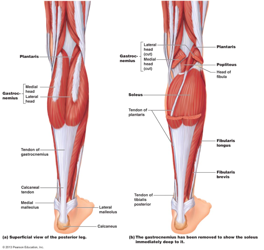
Anatomical diagram of internal organs.
The muscles of the inner thigh whose tendons lead to the pubis bone of your hip are called the hip adductor muscles.

The achilles tendon attaches the muscles of the calves to the bones of the ankle and foot.
The hamstrings refer to 3 long posterior leg muscles, the biceps femoris, semitendinosus, and semimembranosus upper leg muscles and tendons. The hamstrings refer to 3 long posterior leg muscles, the biceps femoris, semitendinosus, and semimembranosus.
0 Comments:
Posting Komentar